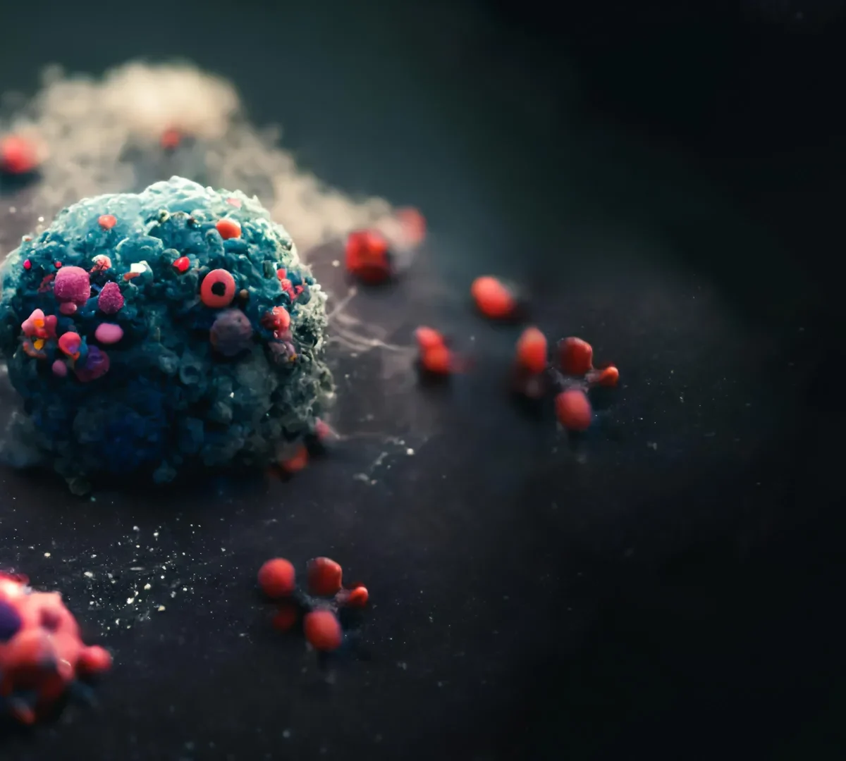
In cities, coworking spaces bring people together to collaborate and innovate. Inside cancer cells, a similar concept plays out — but with deadly consequences. Scientists at the Texas A&M University Health Science Center (Texas A&M Health) have discovered that within the cells of a rare and aggressive kidney cancer, tiny molecular “hubs” form that accelerate disease instead of progress.
Their study, published in Nature Communications, reveals that RNA, typically known for transmitting genetic messages, can be hijacked to build liquid-like “droplet hubs” inside the cell nucleus. These droplets act as command centers that activate growth-related genes. The team not only observed this phenomenon but also developed a molecular switch that can dissolve these hubs on demand, effectively cutting off the cancer’s growth mechanism at its core.
RNA Becomes Cancer’s Builder
The researchers focused on a rare kidney cancer called translocation renal cell carcinoma (tRCC), which primarily affects children and young adults and currently lacks effective treatments. This cancer results from TFE3 oncofusions — abnormal hybrid genes created when chromosomes break and fuse incorrectly.
Until now, scientists did not fully understand how these fusion proteins made tRCC so aggressive. The Texas A&M team found that the fusions enlist RNA to serve as a structural framework. Instead of merely carrying messages, RNA molecules assemble into droplet-like condensates that cluster vital molecules together. These droplets then act as transcriptional hubs, activating genes that promote tumor growth.
“RNA itself is not just a passive messenger, but an active player that helps build these condensates,” said Yun Huang, PhD, professor at the Texas A&M Health Institute of Biosciences and Technology and senior author.
The team also identified an RNA-binding protein called PSPC1, which stabilizes these droplets and makes them even more effective at driving tumor formation.
Mapping the Hidden Machinery of Cancer
To uncover how this process works, the researchers used a suite of cutting-edge molecular biology tools:
- CRISPR gene editing to “tag” fusion proteins in patient-derived cancer cells, letting them track exactly where these proteins go.
- SLAM-seq, a next-generation sequencing method that measures newly made RNA, showing which genes are switched on or off as the droplets form.
- CUT&Tag and RIP-seq to map where the fusion proteins bind DNA and RNA, revealing their precise targets.
- Proteomics to catalog the proteins pulled into the droplets — pinpointing PSPC1 as a key partner.
By layering these techniques, the researchers built the clearest picture yet of how TFE3 oncofusions hijack RNA to build cancer’s growth hubs.
Dissolving the hubs that drive tumors
Discovery alone wasn’t enough. The team wanted to know: If the droplets are cancer’s engine, can we shut them down?
To test this, they engineered a nanobody-based chemogenetic tool — essentially a designer molecular switch. Here’s how it works:
- A nanobody (a miniature antibody fragment) is fused with a dissolver protein.
- The nanobody locks onto the cancer-driving fusion proteins.
- When activated by a chemical trigger, the dissolver melts the droplets, breaking the hubs apart.
The result? Tumor growth ground to a halt in both lab-grown cancer cells and mouse models.
“This is exciting because tRCC has very few effective treatment options today,” said Yubin Zhou, MD, PhD, professor and director of the Center for Translational Cancer Research. “Targeting condensate formation gives us a brand-new angle to attack the cancer, one that traditional drugs have not addressed. It opens the door to therapies that are much more precise and potentially less toxic.”
Beyond Kidney Cancer: A New Therapeutic Model
For the research team, the most powerful part of the study wasn’t just watching RNA build these hubs but seeing that they could be dismantled.
“By mapping how these fusion proteins interact with RNA and other cellular partners, we are not only explaining why this cancer is so aggressive but also revealing weak spots that can be therapeutically exploited,” said Lei Guo, PhD, research assistant professor at the Institute of Biosciences and Technology.
Because many pediatric cancers are also driven by fusion proteins, the implications extend far beyond tRCC. A tool that can dissolve these condensates could represent a general strategy to cut off cancer’s engine rooms at the source.
Why this matters
tRCC represents nearly 30% of renal cancers in children and adolescents, yet treatment choices remain scarce and outcomes are often poor. This breakthrough provides both an explanation for how the cancer organizes its molecular machinery and a potential way to dismantle it.
“This research highlights the power of fundamental science to generate new hope for young patients facing devastating diseases,” Huang added.
Just as cutting power to a coworking hub halts all activity, dissolving cancer’s “droplet hubs” could stop its ability to grow. By revealing how RNA builds these structures — and by finding a way to take them apart — Texas A&M Health researchers have opened a promising new path toward treating one of the most challenging childhood cancers.


